Selective dorsal rhizotomy (SDR) is a common neurosurgical procedure performed in people with cerebral palsy (CP). It reduces spasticity by selectively cutting abnormal sensory nerve rootlets in the spinal cord. (Think of nerve roots like the roots of a tree, which further subdivide into smaller rootlets). SDR only reduces spasticity, not other types of high tone. It is the only irreversible tone-reducing treatment. The following is an explanation of each term:
- Selective: Only certain abnormal nerve rootlets are cut.
- Dorsal: “Dorsal” refers to the sensory nerve rootlets. The dorsal nerve rootlets, i.e., the sensory nerve rootlets, are cut. (The sensory nerve rootlets are termed “dorsal” because they are located toward the back of the body. The motor nerve rootlets are termed “ventral” because they are toward the front.)
- Rhizotomy: “Rhizo” means “root,” and “otomy” means “to cut into.”
Putting it all together, “selective dorsal rhizotomy” means that certain abnormal, dorsal nerve rootlets are cut.
There are two SDR techniques, the cauda and the conus, named after the level of the spinal cord at which each procedure is performed. The choice of technique is provider-specific but also depends on the patient. The cauda technique is the most common worldwide.
SDR involves the removal of the back of the vertebrae (the lamina) in order to access the spinal cord. This is called a laminectomy. During the operation the sensory (dorsal) nerve roots are dissected into rootlets. The rootlets are individually electrically stimulated to determine whether they trigger a normal or abnormal (spastic) response. If a rootlet triggers an abnormal response, it is cut. If not, it is left alone. The percentage of rootlets cut varies between institutions. The higher the number of rootlets cut, the less “selective” the rhizotomy procedure is, and the greater the risk of inducing weakness.
As with many treatments, SDR is not suitable for every child. The selection criteria for SDR differs between institutions. As an example, characteristics of the “ideal” candidate for SDR at Gillette Children’s Specialty Healthcare include:
- Aged 4 to 7 years.
- GMFCS level I–III.
- Primarily spasticity (as opposed to dystonia) that interferes with function.
- Preterm birth history or injury in the late second or early third trimester of pregnancy.
- Periventricular leukomalacia (PVL) confirmed by neuroimaging. (This is the classic brain injury in people with spastic diplegia.)
- Energy-inefficient gait.
- Satisfactory muscle strength, generally defined as antigravity muscle strength at the hips and knees.
- Fair or good selective motor control at the hips and knees. This means the child should be able to partially isolate joint movement (not move the joint in a complete pattern). This requires sufficient strength and motor control; the child shouldn’t be reliant on increased spasticity for their stability or movement.
- Good ability to cooperate with rehabilitation.
Children who fit all of the selection criteria are rare, so physicians use their judgment to determine whether a particular child is a good candidate for SDR. Your provider will assess whether SDR is suitable for your child based on their specific circumstances. (The thinking behind the ideal age of 4 to 7 years is that gait is relatively stable and it is possible to decrease spasticity while the child is still young enough to learn new motor patterns; secondary contracture development is minimal at this point; and the child is old enough to complete the assessment and cooperate with the rigorous rehabilitation program.)
Children for whom SDR is not recommended include those with a predominance of dystonia, less severe spasticity, established contractures (typically in older children and adolescents), weakness, and poor selective motor control. Though SDR is most effective in childhood, it can sometimes be performed in adolescents and adults.
Gait analysis is very important for evaluating whether SDR is appropriate for a particular child and for assessing outcome after surgery.
SDR only reduces spasticity. Any secondary deformities that are present, including muscle contractures and bone deformities, will remain after SDR. These may be treated by orthopedic surgery. SDR is for tone reduction; orthopedic surgery is for bone alignment and residual muscle tightness. It is important to note that as tone-reducing procedures, SDR does not negate the need for orthopedic surgery—they are complementary procedures. The expectation should be that both tone reduction and orthopedic surgery may be needed. However, tone reduction can allow the orthopedic surgery to focus more on bone realignment than on tendon lengthening.
As with any treatment, prior goal-setting and being able to formally evaluate outcome is essential.
SDR is a major operation, and the better the rehabilitation, the better the outcome is likely to be. Just as the operation itself varies between institutions, different institutions have different rehabilitation protocols post-SDR. A review of rehabilitation protocols post-SDR found that patients undergo intensive PT lasting approximately one year starting in the first days after surgery. Patients remain hospitalized anywhere from six days to six weeks.
As an example, at Gillette, rehabilitation begins three days post-SDR and usually involves a four to six-week hospital stay. The benefit of being an inpatient rather than an outpatient is that it allows the patient to attend twice-daily therapy (PT, OT, and other therapies if necessary) and focus on rehabilitation in this early postoperative period. Whether inpatient or outpatient, the aim is to achieve the intensity of postoperative rehabilitation required to maximize functional outcome. A detailed description of the rehabilitation plan is generally provided wherever SDR is carried out.
Overall, there is good evidence supporting SDR from both short-term and long-term studies (up to 26 years post-SDR). Improvements included reduced spasticity and improved gait and function. Long-term follow-up of SDR indicates that over half of the patients underwent orthopedic surgery in the follow-up years. In their long-term follow-up study (10 to 17 years after SDR), Munger and colleagues (2017) reported that non-SDR patients underwent significantly more orthopedic surgery and anti-spasticity injections than SDR patients.
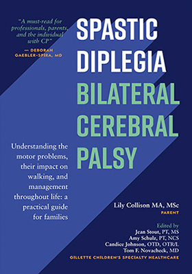
The above is an extract from Spastic Diplegia–Bilateral Cerebral Palsy available here.


